Beyond the Grade: The Embryo Biology Your Clinic's Report Leaves Out

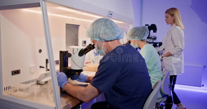
Photo: scientist examining embryo under microscope in fertility lab, via as2.ftcdn.net
When an IVF cycle produces a blastocyst graded 4AA, many patients experience a wave of cautious optimism. The number and letters feel authoritative, almost like a guarantee. Yet embryologists and reproductive endocrinologists know that a grade is, at best, a snapshot—a rough visual estimate of a living structure whose true potential is encoded in layers of biology that no microscope image fully captures.
The gap between what a clinical report communicates and what the science actually measures is significant. Closing that gap, even partially, can transform the way patients engage with their care teams and interpret the outcomes of their cycles.
The Zona Pellucida: An Overlooked Gatekeeper
Surrounding every human embryo is a glycoprotein shell called the zona pellucida. Its primary job during fertilization is to prevent polyspermy—the entry of more than one sperm. But its role does not end there. Throughout the early cleavage stages, the zona provides structural support and influences the timing of hatching, the process by which a blastocyst must break free before it can implant in the uterine lining.
Zona thickness varies between patients and even between embryos within the same cohort. Research published in reproductive medicine journals has explored whether an unusually thick zona may delay or impair hatching, potentially reducing implantation rates. This is partly why assisted hatching—a procedure in which a small opening is made in the zona using a laser or chemical agent—was developed and remains in use at many American fertility centers.
Clinics rarely walk patients through zona measurements or explain why assisted hatching was or was not recommended for their specific embryos. Asking your embryologist about zona characteristics and whether hatching assistance was considered is a reasonable and scientifically grounded question.
Metabolic Markers: What Embryos Exhale
Human embryos consume nutrients and produce metabolic byproducts as they develop. In recent years, researchers have investigated whether analyzing the chemical composition of the culture medium surrounding an embryo—a technique sometimes called metabolomics—can reveal information about viability that morphology alone cannot.
The principle is intuitive: a metabolically active, healthy embryo will interact with its environment in measurable ways. Amino acid turnover, oxygen consumption, and glucose utilization have all been studied as potential markers of developmental competence. While metabolomic profiling has not yet become a standard clinical offering at most US fertility practices, some specialized labs have incorporated oxygen consumption rate measurements as a supplementary assessment tool.
This area of research underscores a larger point: the culture environment itself matters enormously. The specific media formulation a clinic uses, the oxygen concentration in their incubators, and the frequency with which embryos are disturbed for assessment all influence outcomes in ways that never appear on a patient's report.
The Mitochondrial Factor
Mitochondria are the energy-producing organelles within cells, and in the context of embryology, they carry particular significance. Egg cells contain a far greater number of mitochondria than virtually any other human cell type—a reflection of the enormous energy demands of early development. The quality and quantity of mitochondrial DNA within an oocyte, and subsequently within the embryo, has been linked to developmental potential in multiple studies.
Some laboratories have developed mitochondrial DNA (mtDNA) scoring as an adjunct to preimplantation genetic testing. The hypothesis is that embryos with abnormally high mtDNA copy numbers may reflect cellular stress and could have lower implantation rates even when chromosomally normal. The clinical utility of mtDNA scoring remains debated, and it is not universally offered or endorsed. However, if your clinic uses a PGT laboratory that provides this data, understanding what the score means—and what its limitations are—is worth discussing with your physician.
Fragmentation: More Nuanced Than It Appears
Fragmentation—the presence of small, anucleate cellular debris within a developing embryo—is routinely noted on embryo reports and typically influences the grade assigned. High fragmentation is generally associated with poorer outcomes, but the relationship is not linear or absolute.
The location of fragmentation within the embryo, whether it is focal or distributed, and whether fragments are subsequently absorbed or resorbed by the developing cells all affect how meaningfully fragmentation predicts implantation potential. Some research suggests that embryos with moderate fragmentation can self-correct during compaction and blastocyst formation, particularly when the inner cell mass—the cluster of cells that will become the fetus—develops robustly.
Asking your embryologist not just how much fragmentation was observed, but where it was located and whether the embryo showed signs of self-correction, can yield a more textured understanding of a given embryo's prospects.
Time-Lapse Imaging and Kinetic Data
Time-lapse incubation systems, such as those marketed under names like EmbryoScope and Miri TL, capture images of developing embryos at regular intervals without removing them from the incubator. The result is a continuous developmental record that reveals not only morphology at a given moment but the timing and sequence of cell divisions.
Kinetic parameters—such as the precise timing of the transition from two to four cells, or the duration of compaction—have been correlated with implantation outcomes in observational studies. These data points allow embryologists to flag embryos that divided in atypical patterns, even if their final morphology appeared acceptable.
Not all US clinics use time-lapse systems, and among those that do, the degree to which kinetic data informs transfer decisions varies. If your clinic offers time-lapse monitoring, ask whether kinetic algorithms were applied to rank your embryos and what those rankings indicated.
Asking Better Questions
None of this information is meant to suggest that patients should become amateur embryologists or second-guess their clinical teams. Rather, it reflects a simple reality: the more precisely you understand what is being measured—and what is not—the more productive your conversations with your care team will be.
Consider asking your embryologist or reproductive endocrinologist the following during your next consultation:
- What culture media and incubation conditions does your lab use, and how are they optimized?
- Was assisted hatching considered for my embryos, and if so, on what basis?
- Does your PGT laboratory provide mitochondrial DNA scoring, and how do you interpret it?
- Were time-lapse kinetics used in ranking my embryos for transfer?
These are not adversarial questions. They are the questions that demonstrate engagement with the science and invite your care team to share the fuller picture that standard reports simply do not have room to convey.
Embryo development is one of the most complex biological processes in human physiology. The grade on your report is where the conversation begins—not where it ends.