When Development Stops: Understanding Embryo Arrest and What It Can Tell You About Your Reproductive Health
For many people going through IVF, the embryology updates that arrive each morning during a cycle carry enormous emotional weight. A fertilization report brings cautious optimism. A Day 3 check can shift the mood entirely. And when a nurse calls to say that one or more embryos have "arrested" — stopped developing — the news often lands with a particular kind of confusion. The embryo was alive. It fertilized. And then, without explanation, it stopped.
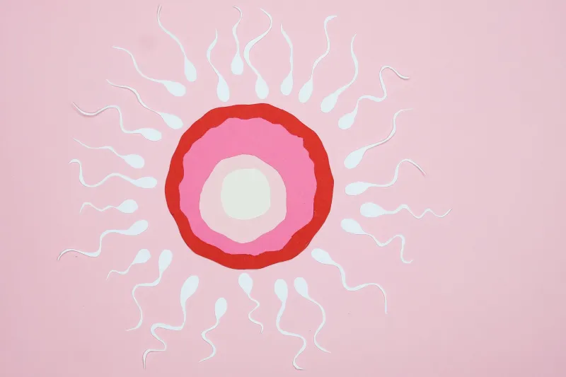
Embryo arrest is not rare. Depending on a patient's age, egg quality, and sperm parameters, a meaningful proportion of fertilized eggs may fail to reach the blastocyst stage by Day 5 or 6. Yet despite how commonly it occurs, it remains one of the least well-explained phenomena in clinical embryology. Understanding the biology behind developmental arrest — and what different arrest patterns may or may not signal — is an important part of becoming an informed participant in your own care.
What Embryo Arrest Actually Means
After fertilization, a human embryo undergoes a tightly choreographed sequence of cell divisions. By Day 2, a healthy embryo typically contains 2 to 4 cells. By Day 3, it may reach 6 to 8 cells. Between Days 4 and 5, the embryo compacts into a morula, then cavitates into a blastocyst — a structure with a fluid-filled cavity, an inner cell mass that will become the fetus, and an outer layer of cells that will form the placenta.
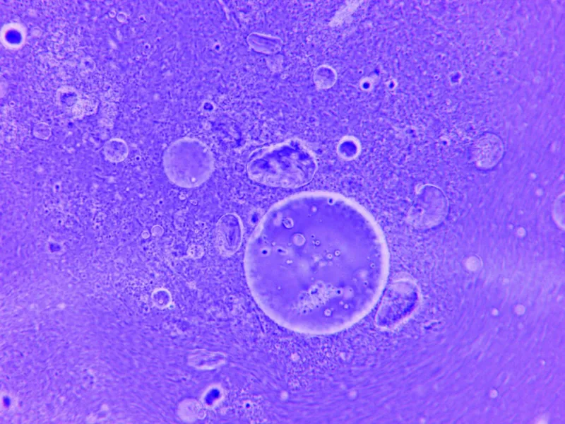
Arrest occurs when this progression stalls. The embryo may stop dividing entirely, or it may fragment to a degree that makes further development impossible. Some embryos arrest early — at the 2- or 4-cell stage on Day 2 or 3. Others reach the morula stage but fail to cavitate into a blastocyst. Still others begin to form a blastocyst but collapse before they can be assessed or vitrified.
Each of these patterns carries different implications, though none provides a simple, definitive diagnosis on its own.
The Biology Behind Developmental Blocks
The earliest stages of embryo development are governed almost entirely by maternal factors — proteins and messenger RNA stored in the egg long before fertilization occurs. The embryo's own genome does not meaningfully activate until what embryologists call embryonic genome activation (EGA), which occurs primarily between the 4- and 8-cell stage in humans, typically around Day 3.
This transition is a critical vulnerability point. If the egg's stored machinery is compromised — due to age-related changes, mitochondrial dysfunction, or abnormalities in how the egg matured — the embryo may not survive the handoff from maternal to embryonic control. Arrest at the 2- to 4-cell stage frequently reflects problems originating in the egg itself, including errors in chromosomal segregation that occurred during meiosis.
Later arrest, between Day 3 and the blastocyst stage, often involves the embryo's own newly activated genome. Chromosomal aneuploidy — having the wrong number of chromosomes — is a leading cause of developmental failure at this stage. Aneuploid embryos frequently arrest rather than progress, which is one reason preimplantation genetic testing for aneuploidy (PGT-A) was developed: to identify embryos with the correct chromosomal complement before transfer.
Sperm quality also plays a role, though it is frequently underestimated. Beyond the sperm parameters captured by a standard semen analysis — count, motility, and morphology — sperm DNA fragmentation can disrupt early embryo development in ways that are not visible at fertilization. High levels of sperm DNA damage have been associated with increased arrest rates, particularly in the days following fertilization when paternal DNA begins to be actively used.
What Arrest Patterns May Reveal
When arrest occurs across multiple embryos in a single cycle, the pattern itself becomes informative — though rarely conclusive without additional context.
Consistent early arrest, concentrated at the 2- to 4-cell stage, often points toward egg quality issues, which may be related to age, ovarian reserve, or the stimulation protocol used. In younger patients with otherwise normal parameters, early arrest may prompt a closer look at sperm DNA integrity, particularly if fertilization rates were normal but subsequent development was poor.
Arrest concentrated at the Day 3 to Day 5 transition — particularly when embryos compact but fail to cavitate — can reflect a mixture of chromosomal abnormalities, mitochondrial insufficiency, or problems with the culture environment in the laboratory itself. This last factor is one reason that outcomes can vary between clinics even when patient characteristics are similar: incubator conditions, culture media, and laboratory protocols all influence how embryos behave in vitro.
A pattern of total fertilization failure or near-total arrest across multiple cycles, even after adjustments to the stimulation protocol, may warrant evaluation for rarer causes, including oocyte activation deficiency — a condition in which eggs fail to activate properly after fertilization and may benefit from assisted oocyte activation techniques during ICSI.
What Clinics Can and Cannot Diagnose From Arrest
It is important to be honest about the limits of what embryo arrest can tell you. In most cases, arrested embryos are not analyzed further. Genetic or molecular testing of arrested embryos is not standard practice, and even when performed in research settings, the results are difficult to interpret because arrested embryos often show complex chromosomal abnormalities that may be artifacts of the arrest itself rather than its cause.
What a clinic can observe is pattern and proportion: how many embryos arrested, at what stage, and whether the pattern is consistent across cycles. What a clinic generally cannot tell you from arrest alone is whether the underlying cause is egg-related, sperm-related, genetic, or environmental — or some combination of all of these.
This ambiguity is frustrating, but it is also an argument for systematic investigation rather than assumption. When arrest is a recurring feature of multiple IVF cycles, a thorough review is warranted — one that includes sperm DNA fragmentation testing, a reassessment of ovarian stimulation protocols, consideration of PGT-A for embryos that do reach the blastocyst stage, and in some cases, a consultation with a reproductive endocrinologist who specializes in recurrent IVF failure.
When to Ask for More Answers
A single cycle with embryo arrest, particularly in patients over 37 or 38, is not automatically a signal that something is wrong beyond the expected challenges of age-related egg quality decline. Chromosomal abnormalities in eggs increase significantly with maternal age, and arrest is often the biological outcome of that reality.
However, arrest affecting the majority of fertilized eggs in a patient under 35, or recurring across two or more cycles despite protocol adjustments, is a reasonable threshold for requesting a more structured conversation with your care team. Questions worth raising include whether sperm DNA fragmentation has been assessed, whether the laboratory's blastulation rates for patients in your age group are consistent with published benchmarks, and whether alternative stimulation approaches — such as a modified natural cycle or a different gonadotropin protocol — might produce a different cohort of eggs.
Embryo arrest is, in the end, a piece of biological information. It is not a verdict on your fertility, and it is not always explainable with current diagnostic tools. But approached carefully and systematically, the patterns it reveals can guide meaningful next steps — and help ensure that each subsequent cycle is informed by what the last one taught you.